Published on
Blood Clot Management: Exploring the EKOS Catheter
People who develop a blood clot in the leg veins (called deep vein thrombosis or DVT) or lung arteries (called pulmonary embolism or PE) need urgent treatment with blood-thinning drugs (called anticoagulants). These medications prevent new blood clots from forming and are usually effective in preventing existing blood clots from growing or moving to the lung arteries. But even with blood-thinning drugs, patients with large or extensive blood clots are still at risk for serious health problems. Patients with PE may experience a worsening of their condition that leads to long-term disability or death. Patients with DVT may continue to struggle with chronic leg pain, swelling, and skin breakdown, reducing their activity and quality of life.
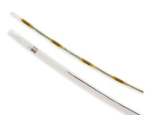
For these reasons, several catheter-based treatments have been developed to help rapidly remove blood clots. These treatments are given with blood-thinning medications and administered by doctors with specialized training, such as interventional cardiologists, interventional radiologists, or vascular surgeons. One such catheter-based treatment known as “ultrasound-assisted thrombolysis” (UAT) involves the EKOS™ Catheter, a device that’s designed to: (1) deliver a clot-busting drug to dissolve existing clots; and (2) use energy from sound waves to spread the drug out within the clot.
TREATMENT OF PE
The EKOS Catheter has been evaluated for the treatment of severe PE—meaning patients at imminent risk of death or who are having heart strain. It was approved by the U.S. Food and Drug Administration (FDA) for this purpose in 2014. With this treatment, the doctor uses imaging with ultrasound and x-rays to see the blood clots and position the EKOS Catheter within the clots through a small skin incision. A low dose of clot-busting drug is then infused through the catheter along with sound wave energy for 6-24 hours. During this time, the patient is monitored carefully in an intensive care unit. After the treatment is completed, the catheter is removed, and compression is applied to the site for about 10-15 minutes to help the small vein incision close.
Among the different methods that can be used to remove blood clots in patients with PE, EKOS Catheter UAT is currently the best-studied method. Key findings from studies include the following:
| Randomized Trial | Who Was Studied | Findings |
|---|---|---|
| SEATTLE II | 150 patients with PE that was severe enough to cause right heart enlargement | Patients who received EKOS Catheter UAT along with blood-thinning drugs (anticoagulants) experienced substantial clot removal and recovery of more normal heart size compared to before treatment. About 10% of patients experienced bleeding with the clot-busting drugs. |
| OPTALYSE | 100 patients with PE | Found that with EKOS Catheter UAT and blood-thinning drugs, reduction in abnormal right heart enlargement can be achieved using very small doses of clot-busting drug (which may improve safety). |
| ULTIMA | 50 patients with PE who were receiving blood-thinning drugs | Improvement in right heart enlargement was significantly greater in patients who were also treated with EKOS Catheter UAT versus patients who were not. This study is the only completed randomized trial comparing catheter-based treatments for PE with standard blood-thinning therapy. |
A larger trial (HI-PEITHO) is now underway. This trial is large (about 500 patients) and is designed to confidently determine if EKOS Catheter UAT will improve important PE patient outcomes with reasonable safety. If so, then it would be reasonable to offer the treatment to more patients.
TREATMENT OF DVT
EKOS Catheter UAT has also been used to treat patients with extensive DVT. Some doctors have adopted this method as their go-to treatment in hopes of reducing leg pain, swelling, and late post-thrombotic syndrome to a greater degree than other treatments. However, more research is needed to determine if that is truly the case.

BostonScientific.com
To date, one small pilot randomized trial of 50 patients (the BERNUTIFUL trial) confirmed that use of the EKOS Catheter (with or without the sound wave energy) to deliver clot-busting drugs removed clots from the leg veins – but benefits weren’t associated with the sound wave energy itself.
Another randomized trial of 184 patients (the CAVA trial) found EKOS Catheter UAT to successfully remove blood clots; however, it didn’t result in fewer leg symptoms or better quality of life than blood-thinning drugs alone.
The role of EKOS Catheter UAT for DVT continues to be discussed and studied. While catheter-based therapies appear to be helpful for some patients with the most extensive blood clots, the best method to use is not clear at this time.
THE BOTTOM LINE
In recent years, science and innovation have produced new blood-thinning drugs, new imaging methods, and new catheter-based treatments with the potential to help patients with PE and DVT. Early studies suggest that EKOS Catheter UAT can rapidly remove clots in both conditions. Additional studies will help patients and doctors understand how to best translate such tools into clinical benefits for patients, and which patients should be treated.
*This article is for informational purposes only. NATF does not endorse any specific type of catheter-directed treatment or any manufacturer of such treatments.
GLOSSARY
| Term | Definition |
|---|---|
| Interventional cardiologist | A doctor that specializes in delivering minimally invasive treatments for diseases of the heart or other blood vessels. |
| Interventional radiologist | A doctor that specializes in delivering imaging-guided, minimally invasive, catheter-based treatments. |
| Post-thrombotic syndrome | Chronic leg pain after a blood clot. |
| Randomized controlled study | A study in which participants are randomly assigned to separate groups that compare different treatments or interventions. |
| Right heart enlargement | The right side of the heart takes blood from the body and pumps it to the lungs. When a large blood clot moves into the lung arteries (pulmonary embolism or PE), it sometimes causes the right side of the heart to pump against more resistance, which can affect its ability to work properly. When this happens, the right heart often gets bigger. In a patient with PE, right heart enlargement can be a sign of a heart that is not working normally. |
| Ultrasound | Energy from sound waves, which can be used either to image the body or, in a different form, to spread clot-busting drug within blood clots. |
| Vascular surgeon | A doctor that specializes in doing surgery or minimally invasive treatments on blood vessels, including veins. |

This article was authored by:
Suresh Vedantham, MD
Assistant Dean for Clinical Research
Professor of Radiology and Surgery
Washington University School of Medicine


